Cells
You should be able to:
Identify three major cell components and their structures/functions.
Learn about methods of transport inside and outside of cells.
Examine in detail the function of the nucleus.
What are cells?
Cells are the smallest units of life within the human body. There are roughly over 100 trillion cells in the human body with 200 different functions.¹ Cells are often specialized depending on their overall function, composing what is known as tissues .¹ Organs are composed of two or more tissues with similar functions and identifiable shapes.¹
Three Major Cell Structures
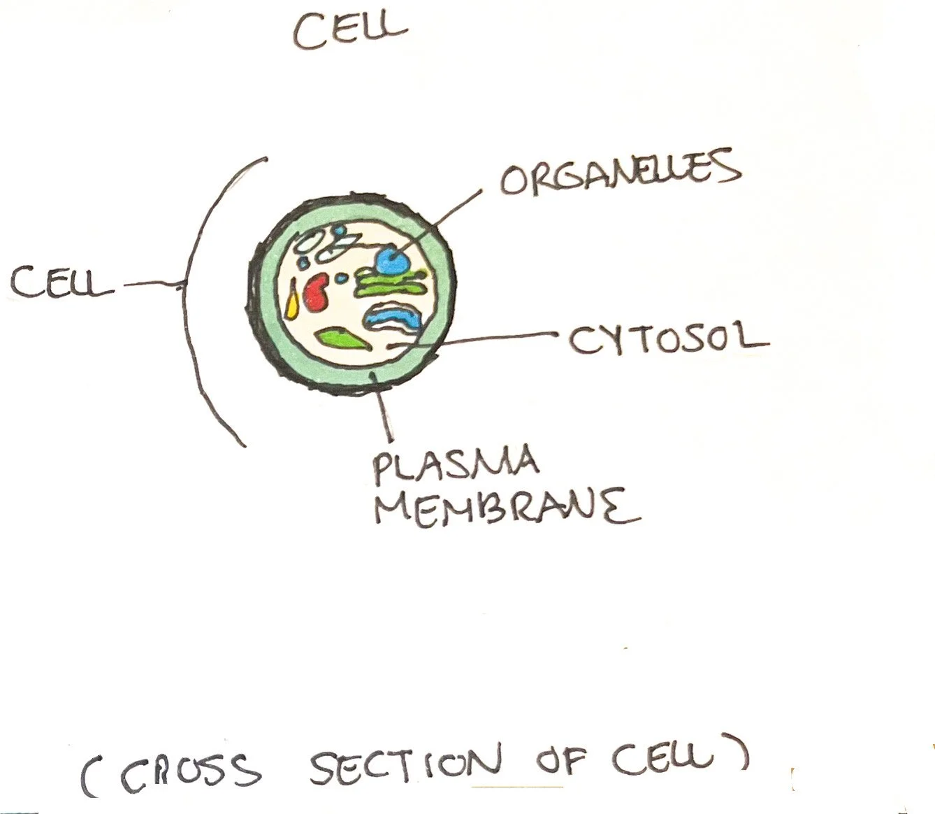
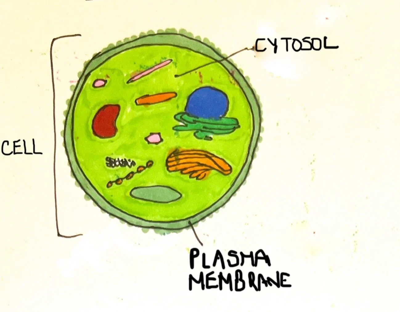
To understand cells, it is important to understand the composition in and around them. Cells have the following structures from the outermost to the innermost part of cells:
Plasma membrane: fluid-like barrier that selectively permits molecules allowing for entrance into the cell or exit from the cell.
Cytoplasm: inside the cell contains cytosol, also known as intracellular fluid.
Organelles: Organ-like matter that has specific functions to sustain life.
Plasma Membrane
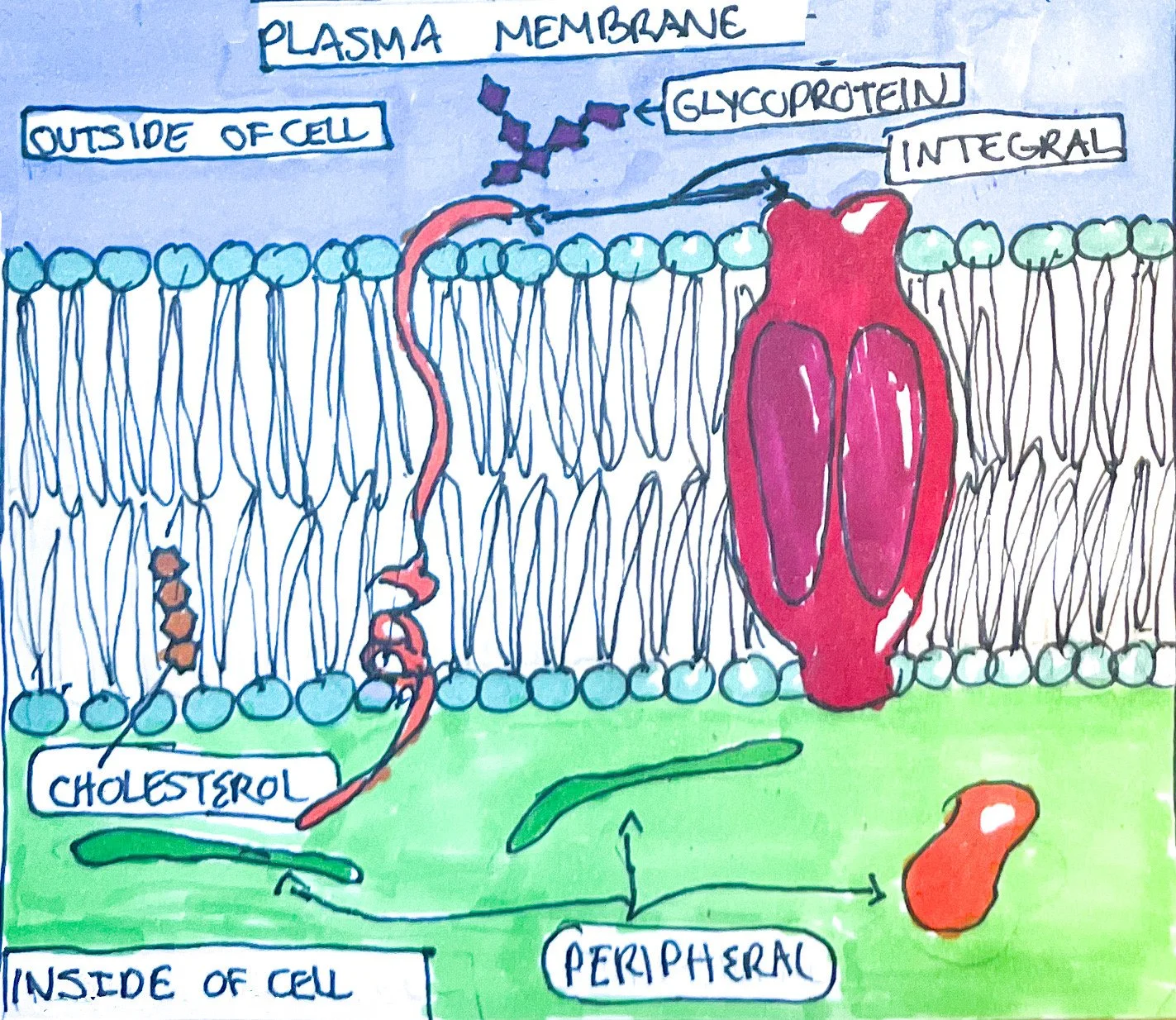
Structure: The structure of the plasma membrane, separating the inside and outside of the cell, consists of a lipid bilayer. The lipid bilayer is structured where the polar/water soluble heads are directed into the extracellular fluid and the cytosol. The middle is home to the nonpolar/fat-soluble tails. Cholesterol (steroid with hydroxyl group) and glycolipids (lipids with carbohydrate component) are present within the plasma membrane as well.¹
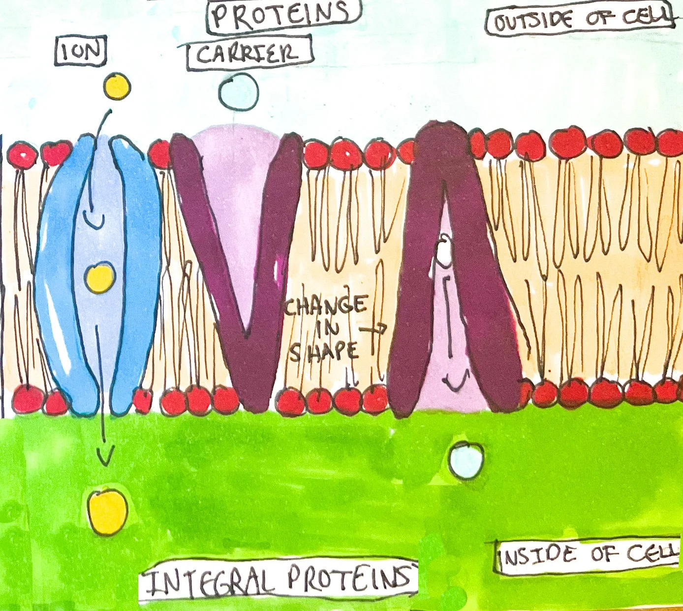
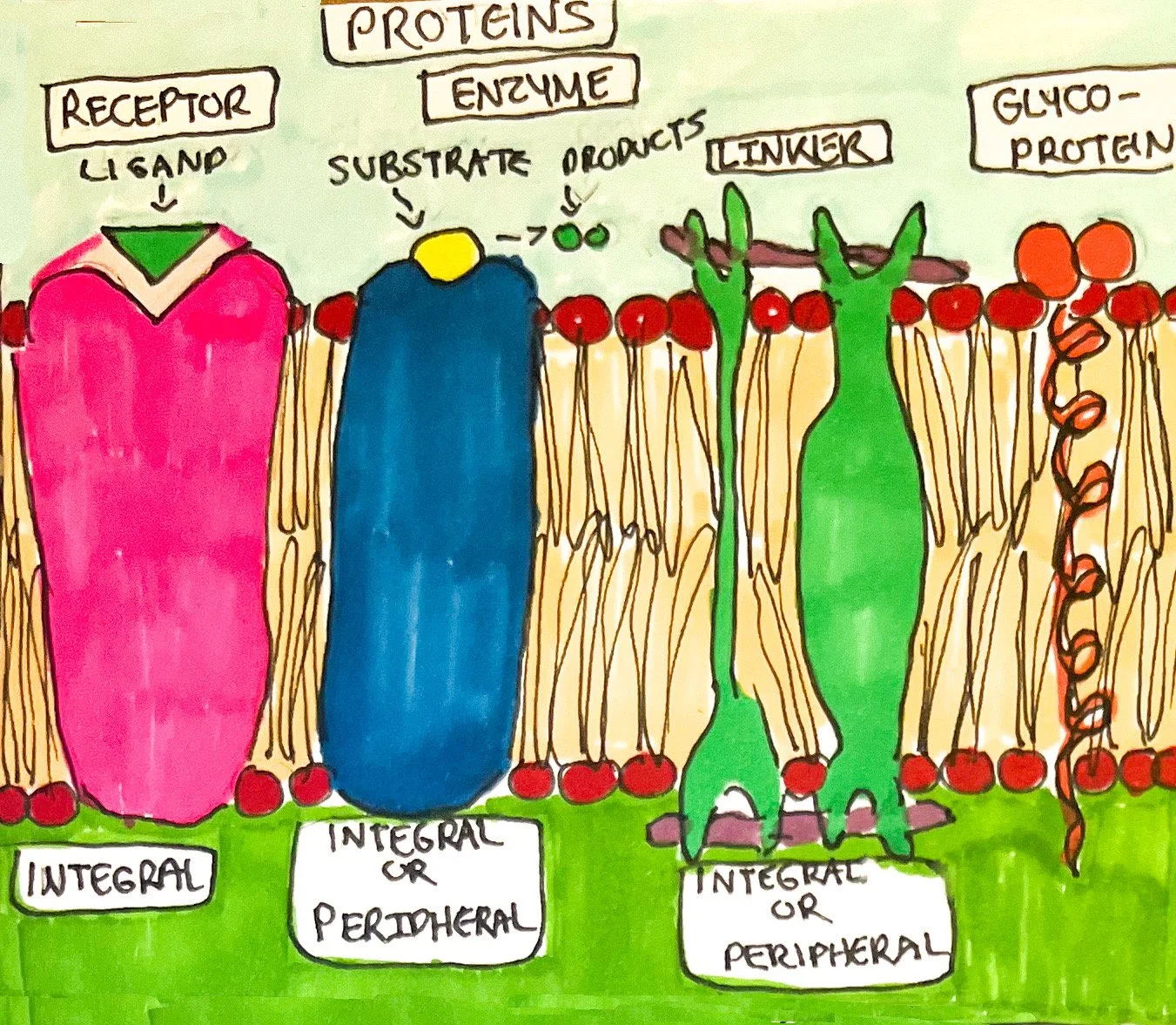
Proteins: Plasma membrane proteins are also part of the structure. Transmembrane proteins extend from the cytosol to the extracellular fluid through the plasma membrane.¹ These proteins are known as integral proteins and are embedded within the membrane.¹ Glycoproteins (proteins with carbohydrate components) are a common type of integral protein that forms the glycocalyx of cells.¹ The glycocalyx is an identifier for cells since the carbohydrate component forms a unique pattern for each cell, aiding in cell recognition and movement.¹ Peripheral proteins are not embedded within the plasma membrane and move more freely around the polar heads of the membrane or on integral proteins.¹
Function of Proteins: The plasma membrane functions as a selective permeability barrier and has many integral and peripheral proteins whose variety determines the function of cells:
Ion channel (integral): a specific ion can flow across the plasma membrane
Carrier/Transporters(integral): transports specific ions or polar molecules in and out of the cell through changing shape ¹
Receptor (integral): recognizes and binds a specific molecule termed ligand¹
Enzyme (integral and peripheral): catalyze reactions either inside or outside of cells¹
Linker (integral and peripheral): binds cells together and holds filaments to stabilize cell shape and structure¹
Glycoprotein/cell identity marker (integral): identifies cells during tissue creation and invading cells¹
Key Concepts: Diffusion, Osmosis, Active Transport, and Vesicle Transport
The structure of the plasma membrane allows nonpolar or uncharged molecules to pass through the nonpolar, fat-soluble tails of the lipid bilayer.¹ The plasma membrane transports uncharged, nonpolar molecules such as oxygen, carbon dioxide, and steroids more easily than polar, charged molecules. Some small polar molecules, such as water, are permeable.¹
Diffusion
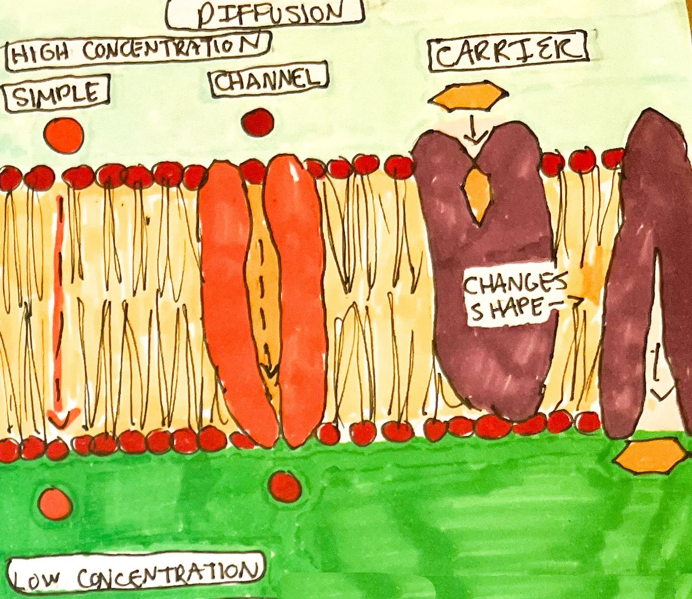
Diffusion: The principle of diffusion demonstrates that when mixing a solution, the solutes (dissolved substances) will move from an area of high concentration to low concentration until equally dispersed through the solvent (dissolving fluid).¹The solutes’ movement from high to low concentration is termed going down the concentration gradient (separate areas that contain differing amounts of solute). The ability of a solute to disperse is dependent on multiple factors. Diffusion is applied to human biology as molecules move in and out of cells to reach and maintain an equilibrium or homeostasis. It is important to note that the overall charge of the concentration gradient is negative inside the cell due to the greater loss of potassium cations.¹ Therefore, the outside of the cell is more positively charged. This is called the electrochemical gradient. Diffusion is considered passive as it does not require more than its own kinetic energy to move across the plasma membrane.
Simple Diffusion: Movement of molecules through the plasma membrane into the cell using its own kinetic energy without plasma proteins.¹ Molecules include fat-soluble vitamins (A, D, E, and K), oxygen, carbon dioxide, water, urea, fatty acids, steroids, etc. Simple diffusion favors nonpolar, uncharged molecules due to the lipid bilayer.
Facilitated Diffusion: Polar, highly charged molecules require the help of plasma proteins to move through plasma membranes into cells. Facilitated diffusion can be either:
Channel-Mediated: Substance passes through channel protein into the cell. Channel proteins are used for ions, especially potassium and chloride ions to go into the cell (less for sodium and calcium).¹
Carrier-Mediated: Substance binds to a carrier protein and passes into the cell as the protein changes shape.¹
Osmosis
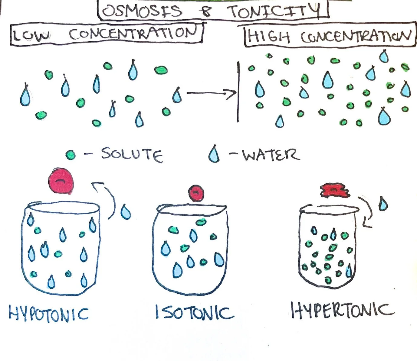
Osmosis: Osmosis, similar to diffusion, is a passive process in that movement depends on concentration. Water moves from an area of high water concentration to a low water concentration.¹ In another sense, water moves from an area of low solute concentration to a high solute concentration. Water moves through simple diffusion or aquaporins (integral plasma membrane channel proteins) that allow for water permeability. Hydrostatic pressure measures the pressure exerted by water against the membrane, and osmotic pressure measures the pressure applied by the solute that is impermeable to the membrane.¹
When a solution is isotonic, the osmotic pressure is at an equilibrium. A hypotonic solution contains more water than solute, so water moves from high water concentration to low water concentration inside of the cell which can cause swelling and bursting.¹ A hypertonic solution contains less water compared to solute, and water from cells exits into extracellular fluid, causing shrinkage (crenation) of cells.¹
Active Transport
Unlike diffusion, active transport requires energy either in the form of adenosine triphosphate or energy provided by the electrical charge of the concentration gradient. Active transport moves monosaccharides, amino acids, and ions such as sodium, potassium, hydrogen, calcium, iodide, and chloride against the concentration gradient or uphill; however, they are also transported by facilitated diffusion at times.¹ There are two types of active transport:
Primary Active Transport: The action of hydrolysis of ATP releases phosphate which changes the carrier protein shape once attached, allowing one substance to travel against the concentration gradient. The other substance crosses once the phosphate is removed from the carrier protein.¹
The sodium-potassium pump is one of the most common forms of primary active transport found within the body with each bringing two potassium cations into the cytoplasm and three sodium cations into the extracellular fluid.¹ Therefore, the outside of the cell is more positively charged than the inside of the cell.
Secondary Active Transport: Solutes are transported against the concentration gradient at the same time either in the same direction (symporter) or opposite direction (antiporter) due to the leakage of sodium back into the cell.¹ The constant motion of expelling sodium maintains the concentration gradient and allows for sodium-calcium and sodium-hydrogen antiporters and sodium-glucose and sodium-glucose symporters to function.
Vesicles
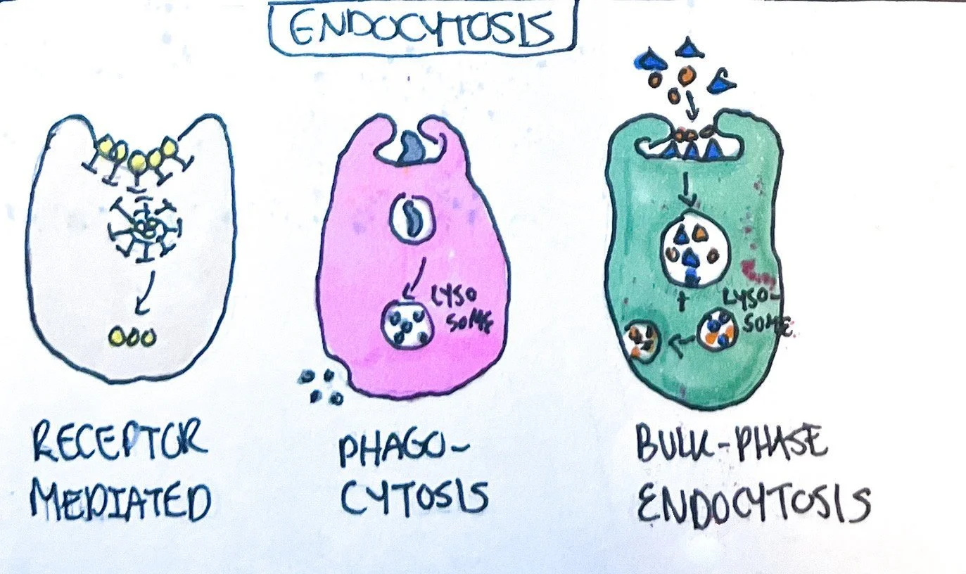
Vesicles are sacs formed within the plasma membrane that transport substances into cells (endocytosis) and out of cells (exocytosis). There are three types of endocytosis:
Endocytosis
Receptor-mediated endocytosis: Once a substance (ligand) binds to the extracellular side of the plasma membrane via receptor proteins, a vesicle forms and transports the substance to organelles. Substances transported via endocytosis include low-density lipoprotein (LDL) cholesterol, vitamins, and certain hormones.¹
Phagocytosis: Phagocyte cells, such as macrophages and neutrophils, engulf microbes and other substances once bonded to a receptor protein, forming the vesicle termed a phagosome that undergoes processes for degradation within the lysosome and excretion of residue.¹
Bulk-phase endocytosis: Solutes within the extracellular fluid are transported into the cell without the use of a receptor protein when vesicles form within the plasma membrane and pinch off to combine with a lysosome.¹
Exocytosis
Secretory vesicles form within the cell on the innermost part of the plasma membrane and release substances outside the cell into the extracellular fluid.¹ This is very important for cells that release secretions and neurotransmitters to get rid of waste.

2. Cytoplasm
The cytoplasm is composed of the cytosol (or the intracellular fluid), and organelles (small organ-like structures allowing cells to function).¹
Cytosol: The cytosol ranges between 75-90% water with suspended ions, amino acids, glucose, fatty acids, etc.¹ The cytosol has a cytoskeleton composed of microfilaments, intermediate filaments, and microtubules that aid in cell structure and movement.
3. Organelles
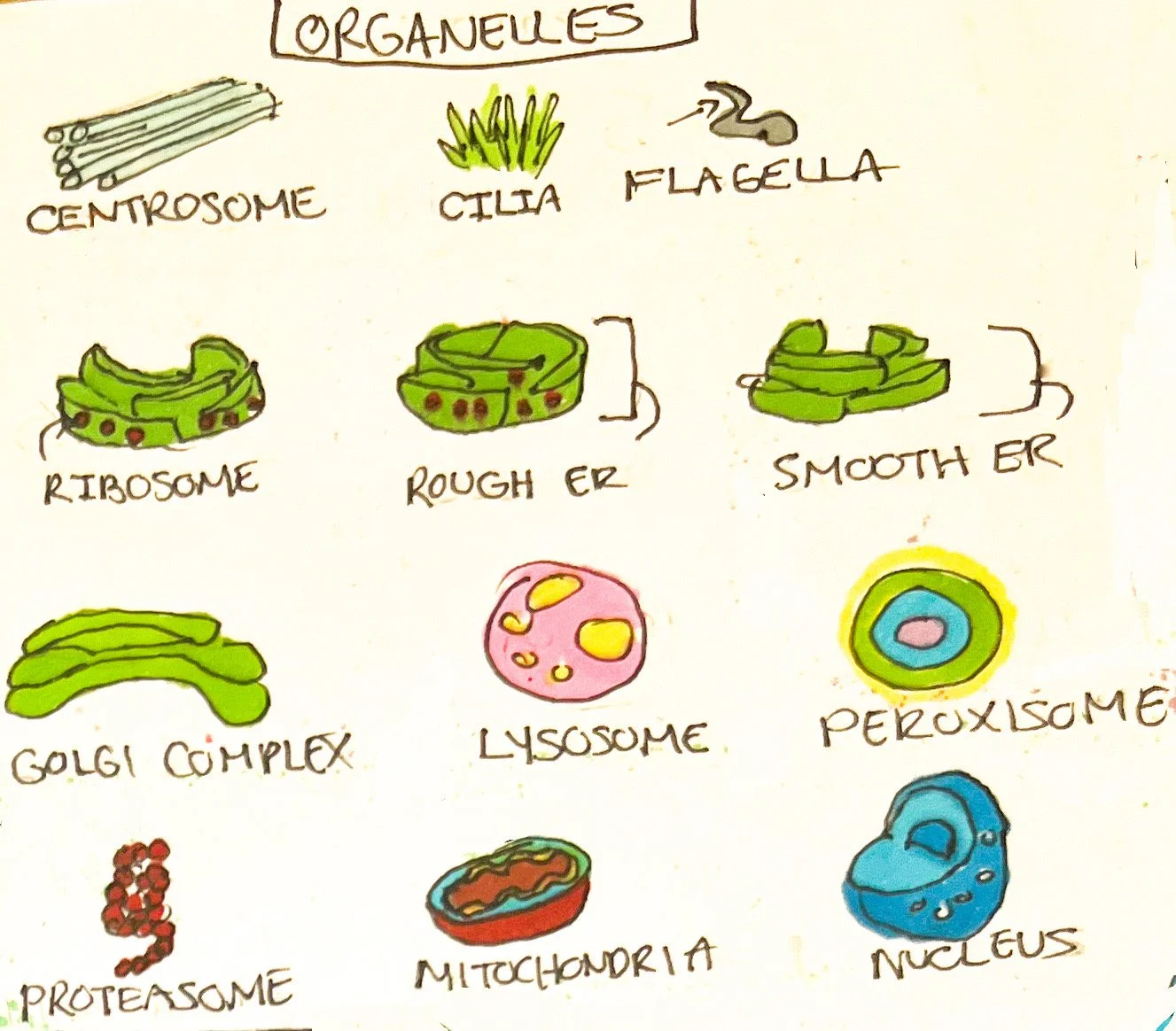
Organelles function to maintain cellular life. They are present in varying amounts within cells depending on the function of the cell. The following are types of organelles and their functions:
Centrosome: Aids in building microtubules in cells that are unable to divide, and a mitotic spindle is needed during cell division.¹
Cilia and Flagella: Cilia are microprojections on the cell surface helping in substance movement while flagella move the cell as a whole.¹
Ribosomes: site for protein synthesis composed of ribonucleic acid (RNA). ¹
Endoplasmic Reticulum: Tubules that come in two varieties:
Rough ER: tubule is studded with ribosomes. Once proteins are synthesized, they pass through the rough ER to become secretory, membrane, and organelle proteins.¹
Smooth ER: Synthesizes fatty acids and steroids, detoxifies substances, and allows substances to be released.¹
Golgi Complex: modifies and transports proteins from rough ER and forms membrane, secretory, and transport vesicles.¹
Lysosomes: vesicles that digest substances and perform autophagy (organelle digestion), autolysis (destruction of the whole cell), and extracellular digestion.¹
Peroxisomes: oxidizes organic and toxic substances such as hydrogen peroxide via enzymes.¹
Proteasomes: destroys cytosolic proteins with enzymes serving as negative feedback for metabolic processes.¹
Mitochondria: Powerhouse of the cell that generates adenosine triphosphate (ATP) and signals for apoptosis (programmed cell death).¹
Nucleus: Largest organelle within the cell that has pores controlling the movement of substances, nucleoli that create ribosomes, and chromosomes that control the structure and function of cells.¹
Processes within the Nucleus
Protein Synthesis
Transcription: Chromosomes house genes that contain templates for protein synthesis. During transcription, messenger RNA (mRNA), ribosomal DNA (rRNA), and transfer RNA (tRNA) are created from deoxyribonucleic acid (DNA) within the nucleus.¹
Translation: During translation, the mRNA attaches to the ribosome and tRNA binds to a specific site on the ribosome to add anticodons. This allows for amino acids on the tRNA to form peptide bonds and create a chain of polypeptides coding for a protein.¹
DNA synthesis
Similar to protein synthesis but occurs solely within the nucleus. DNA strand is split by enzymes and RNA nucleotides begin the process in which DNA polymerase adds corresponding DNA nucleotides.¹ RNA nucleotides are removed by an enzyme to allow DNA polymerase to add DNA and seal into a continuous strand by another enzyme.¹
Cell Synthesis
Interphase: This occurs prior to mitosis and meiosis.¹Interphase consists of G1, S, and G2 phases. Cells that are relatively nondividing, such as nerve cells, are in the G0 phase.¹ G1 phase is when organelles replicate.¹ S phase is where DNA is replicated, and G2 wraps up synthesis of proteins and enzymes, among other structures, needed for the cell to begin mitosis or meiosis.¹
Chromatin/Chromosomes/Chromatids: Chromatin is the precursor of chromosomes that consists of DNA, RNA, and proteins.¹ Chromosomes consist of DNA compressed into one or two chromatids.¹ However, one chromatid is often referred to as being half of the chromosome.¹ A chromatid is held by a centromere in the middle. Chromosomes are counted by the centromeres and not the chromatids.¹
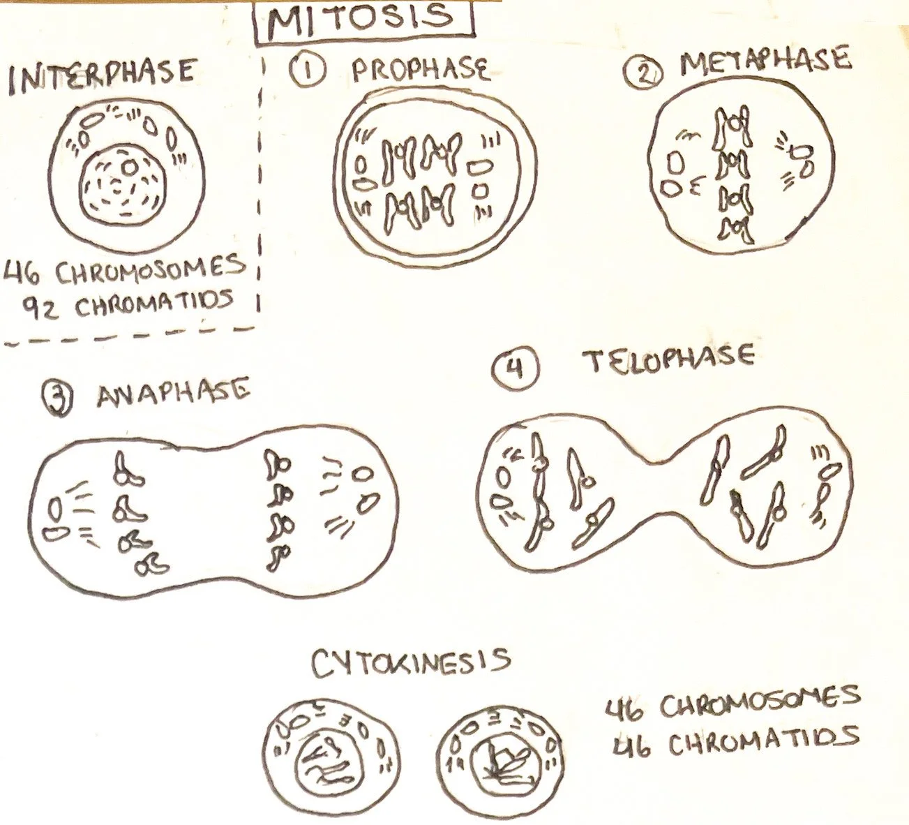
Mitosis
Mitosis is a form of cell division to create two identical cells that contain 23 pairs of chromosomes each (46 chromosomes each), known as diploid cells or somatic (body) cells. Mitosis is best described in phases known as prophase, metaphase, anaphase, and telophase.¹ In prophase, chromatids from interphase are formed into their proper structure with the development of the mitotic spindle used to separate chromatids later. ¹ Metaphase aligns the paired chromatids and anaphase splits the paired chromatids and cleavages the cell into two.¹ In telophase, the chromosomes unravel into chromatin, and the nuclear envelope is formed.
Cytokinesis: During anaphase and through telophase, cytokinesis allows for the equal splitting of organelles and cytoplasm allows for the cell to be properly configured. Once completed, the cell is split into two diploid cells.¹
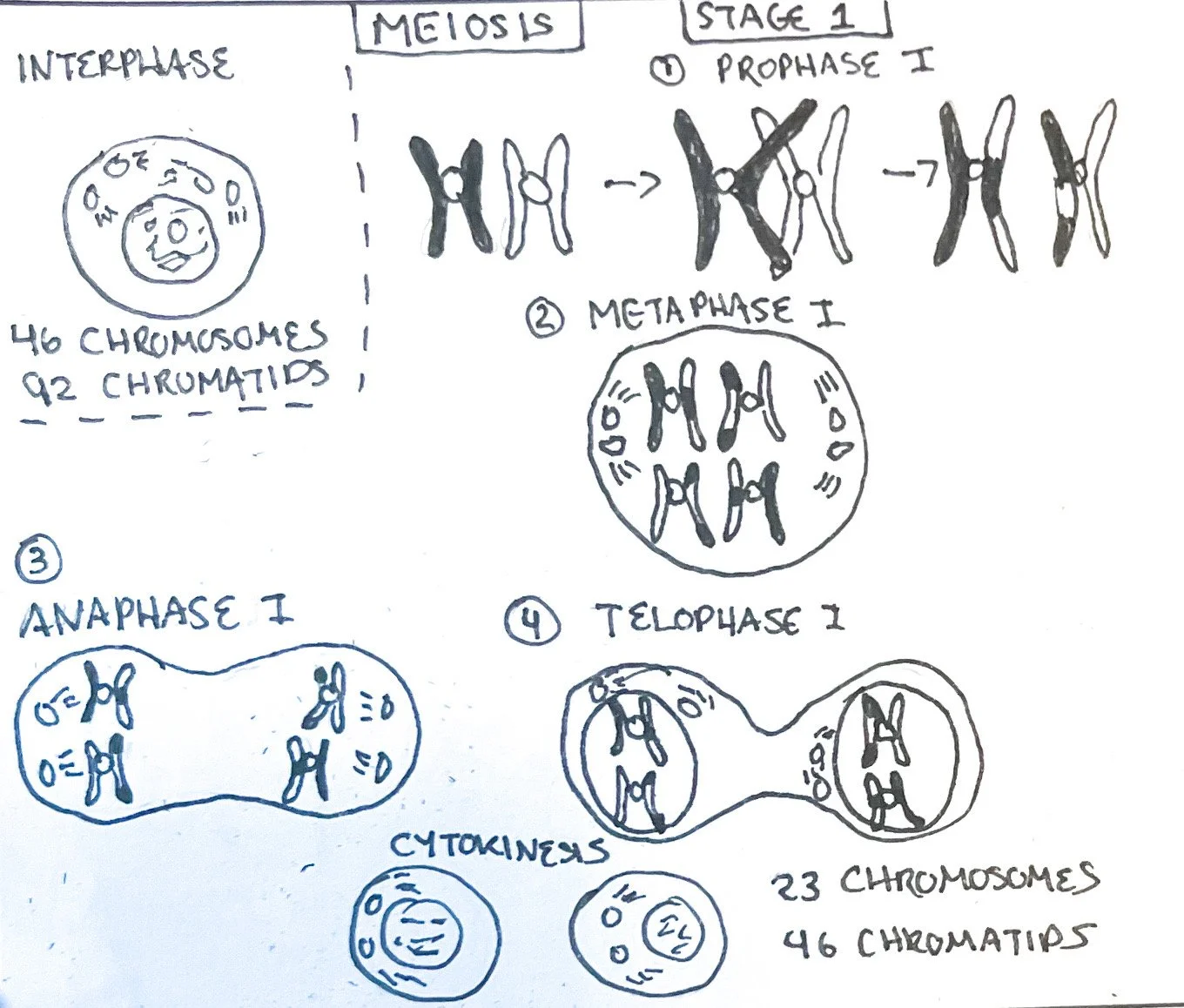
Meiosis
Meiosis is a form of cell division creating four cells that contain 23 chromosomes, known as haploid cells specific to sexual reproduction such as sperm or oocyte.¹ Meiosis is divided into two stages called meiosis I and II that contain the prophase, metaphase, anaphase, and telophase similar to mitosis (categorized as I or II depending on the stage).¹ Before the phases, interphase occurs similar to mitosis. This means that a diploid cell (46 chromosomes) is duplicated to have 46 chromosomes with 92 chromatids.¹
Meiosis I: Beginning with prophase I, the sister chromatids (arising from replication and genetically identical)are formed into tetrads (a group of two pairs of chromosomes containing two chromatids) in a process called synapsis.¹ They may cross over, allowing for non-sister chromatids to share genetic info and allow for genetic differences among humans.¹ Metaphase I aligns tetrads, and anaphase I separates homologous chromatids with sister chromatids remaining together.¹ Telophase I cleavages the cell in two and cytokinesis occurs, resulting in two haploid cells with 23 chromosomes and 46 chromatids.¹
Meiosis II: Next, the two haploid cells enter each phase again (termed prophase II, metaphase II, anaphase II, and telophase II), resulting in 4 haploid cells containing 23 chromosomes and 23 chromatids.¹ It is similar to mitosis.
Source(s)
Jerry Tortora and Bryan Dickerson, Principles of Anatomy & Physiology, 16th ed. (New Jersey: John Wiley & Sons, 2021).